3D Scanning a Medical Device

As part of the design course I decided to 3D scan a medical device. 3D scanners can be handheld or stationary and work by cycling vertical or horizontal laser lines on/off the object. A trackable difference between the object and other lights are captured during scanning and two cameras are able to capture the surface geometry of the object based on the pattern distortion. The sensing cameras observe the contrast along the lines edges and assign those pixels as xyz coordinates which match a point on the surface of the object being scanning. The shape of the object appears as millions of points called a “point cloud” on the computer monitor. After the huge point cloud data is created it is registered and merged into one three-dimensional representation of the object through the process of meshing. The meshing process calculates how the points relate to each other in order to join them together into surfaces. 3D scanning is an up and coming technology used today for reverse engineering, creating personalized medical solutions, facial reconstruction in archaeology and forensics and in the future, is being looked into aiding the possibility and improvement of self-driving cars.

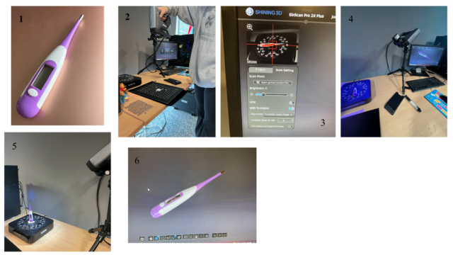
In the first picture you can see the digital thermometer that was used for the scanning. The first crucial step when using a 3D scanner is the calibration, which you can see being done in the second picture. Due to the reliance of light for the entire process, change in lighting can affect the quality of the scan. When calibrating you are allowing the device to calibrate to the current state of the surrounding light. In the third picture you can see the settings that are available before you scan. The brightness helps and determines what the scanner will be able to pick up. When you can see the shape of the object clearly and there are some red dots on the monitor, the brightness is adjusted well for the scan. The red represents extreme contrast. You adjust the brightness depending on the colors of the object being scanned. In the fourth picture you can see the 3D scanner mid scan. It displays purple laser lines vertically and flashes a white light and on the device. The fifth picture shows another position for the thermometer. It is important to get multiple angles when 3D scanning in order to get the best data from the device. The software is able to detect similar geometry and then combine the different scans together in order to create the best representation of the object. The sixth picture is what the final scanned 3D model looks like in the software, which can then be uploaded into a CAD program.

Benefits of Ecosystem Mapping

Before coming to Scotland to take the Honors 352 course, I had never heard the term, “ecosystem mapping”; after five weeks, we are all very well acquainted with this term. During our first week in Dundee, Rod Mountain introduced us to this creative tool used by healthcare professionals and healthcare management teams to understand the patient demographics, service pathways, and national policies in place for their country’s healthcare system. Over the next four weeks, we used this tool in various iterations to better understand and be able to compare the healthcare systems of the United States, Scotland’s NHS, and other European countries (my group focused on the Italian healthcare system).

As part of our final project for the class, we were tasked to use the ecosystem map to create an ideal healthcare system for our region of choice, set to take effect in 2040. My group decided to design a future healthcare system for Italy, having already heard from several Italians we had met about what needed to be changed in their current system. It’s really easy to talk about all the things wrong with a country’s healthcare system, and to idealize how a perfect system would function, but what was challenging about this assignment was that we had to design a realistic healthcare system with policies supporting our design. We had to think critically about the types of people that make up Italy’s population, and tailor a system specifically for that population, keeping in mind aspects of the current system that were working and we wanted to maintain in the future. As a BME major, healthcare policy is more unfamiliar to me than physical design, but it was rewarding to see how interconnected all the branches of healthcare are, including biomedical engineers. The ecosystem mapping exercises were very beneficial to me and really helped focus my thinking while brainstorming about the “ideal” healthcare system, which has changed the way I see and think about healthcare design.


My Designed and Printed Objects!

Creator: E. Nathaniel McBride

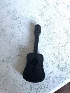
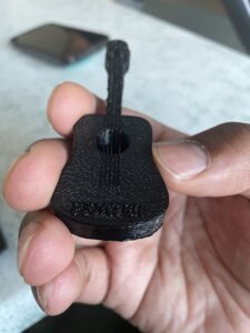
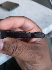
  1. This guitar features pluckable strings and is engraved with Floater, a homage to the friends I made while studying abroad in Scotland.

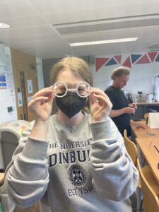
2. I also designed and printed glasses. I received inspiration from Harry Potter. As a result, I named my glasses “Potter” and had my friend Kayley model them.

Dolly the Sheep

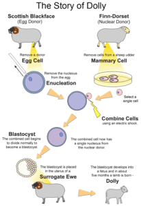
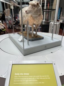
Dolly the sheep was the first mammal to be cloned using biomedical engineering from an adult cell. This experiment took place at the Roslin Institute, at The University of Edinburgh. Professor Sir Ian Wilmut led the research team to clone a cell from a Finn Dorset Sheep, which proved to be successful when Dolly did not share the black markings that her surrogate mother had. 

This experiment created a multitude of possibilities within biology, while also raising several questions concerned with the ethics of cloning (Roslin Institute, n.d.). 

Dolly was cloned using the Somatic Cell Nuclear Transfer, or SCNT. For this, the nucleus of a somatic, body cell is transferred into the cytoplasm of an egg cell that has its nucleus removed. The nucleus from the somatic cell replaces the original nucleus from the egg cell to become a zygote (fertilized egg). With this method, extinct species could be resurrected, such as the wooly mammoth with an elephant surrogate (Stocum & Rogers, 2009). 

Dolly died in 2003, but she is now located in the National Museum of Scotland in Edinburgh. 

The Life of Dolly. (n.d.). Dolly the Sheep. Retrieved July 6, 2022, from https://dolly.roslin.ed.ac.uk/facts/the-life-of-dolly/index.html

Stocum, D., & Rogers, K. (2009). somatic cell nuclear transfer | Definition, Steps, Applications, & Facts. Encyclopedia Britannica. Retrieved July 6, 2022, from https://www.britannica.com/science/somatic-cell-nuclear-transfer 
  

3D Printed Cannon – Christian Nightingale

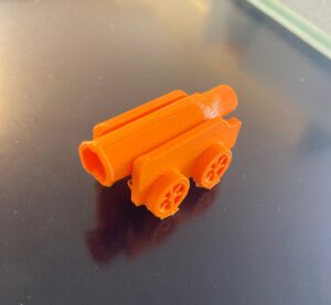
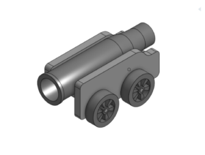
This week, I 3D printed a cannon like the ones that are surrounding Edinburgh Castle. Specifically, this cannon was based on the Mons Meg cannon that was gifted to King James II of England in 1457. The cannon can fire a 150kg cannonball up to 2 miles. I also made a short video that shows the steps taken to 3D print the cannon!


Visiting Surgeon’s Hall Museum

The Surgeon’s Hall Museum in Ediburgh was an amazing experience to see how far science and technology have improved in the past few centuries. The Museum featured a history of surgery exhibition, which, as a pre-med student, was one of my most interesting endeavors on our trip. The exhibit featured tools and techniques used in surgery over the past several centuries. Prior to this trip, I gave little thought to medical products and their innovation. I visited the museum during our last week, and it was perfect timing; I had already learned so much about biomedical engineering and design during the program, which made visiting the Museum in the last week the perfect way to cap my learning. 

The tools and techniques used in the 17th century, and on, are utterly shocking to me. Surgery was performed without anesthesia, without antiseptics, and without clean tools. The Museum walked you through how surgery has evolved, from grave-robbing (which Scotland was notoriously known for) to learn the basis of human anatomy, to keyhole surgeries in the modern day. The Surgeon’s Museum was a great way to not only learn about human anatomy, but also perfect for applying biomedical engineering to healthcare. It helped me, a Biology major, learn more about how important biomedical engineering truly is for improving post-operation outcomes. I would definitely recommend visiting the museum to anyone in Edinburgh!