The Body Voyager Exhibit

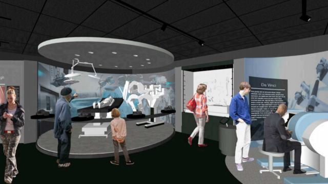
My favorite educational experience on this trip, outside of the classroom, was my visit to the Surgeons’ Hall Museum. My favorite part of the museum was The Body Voyager Gallery which is an immersive exhibit that centers around robotic surgery and technological advancements in the medical field. As a biomedical engineering student, concentrating in robotics, I am obviously very interested in the ways technology is being merged with medicine and healthcare. As this new face of healthcare is being introduced I am very excited and fascinated to see, and take part, in the developments that will be made technologically in the medical field. This experience gave me hands-on experience and expanded my knowledge on the future possibilities in biomedical engineering. I learned about new technology in oral surgery and facial reconstruction, was able to try out what it would be like to perform keyhole surgery by using a laparoscopic instrument to stack blocks through a tiny hole, I was able to paint a picture using a Da Vinci surgical machine and learn about all the parts of a surgical robot.

With healthcare moving in a technological direction this exhibit helped open my eyes about all the changes that are going to take place in the design of healthcare. Technology is changing the course of surgery and patient care and people are going to have to make, design, improve, fix, manage and be able to use all of this new technology. Healthcare will need knowledge and aid from a plethora of different fields. In order to be successful, input is going to be needed not just from engineers and doctors but from designers, policy makers, teachers and so much more. The ideas for these advancements will need to be brought to life, built, implemented, managed and taught how to be used. New people will be needed to fix mechanical problems with the machines and be able to teach people how to use the machines properly. Integrating technology with healthcare will present numerous opportunities for improving and transforming healthcare design. 

Sheep, Sheep and More Sheep

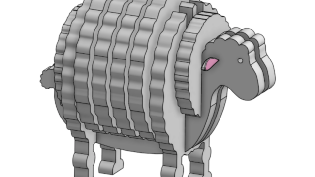
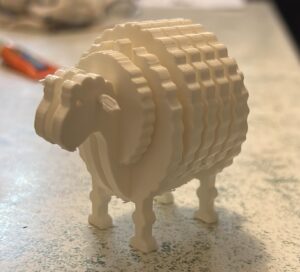
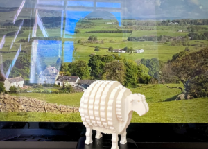
When tasked with designing an object that spoke to my personal experience in Scotland I could only think of one thing. From my time spent in Scotland I have traveled quite a bit across the entire country. When traveling between different locations I could always count on seeing lots and lots of sheep out my window, which makes sense as Scotland is known for having more sheep than people.

3D Scanning a Medical Device

As part of the design course I decided to 3D scan a medical device. 3D scanners can be handheld or stationary and work by cycling vertical or horizontal laser lines on/off the object. A trackable difference between the object and other lights are captured during scanning and two cameras are able to capture the surface geometry of the object based on the pattern distortion. The sensing cameras observe the contrast along the lines edges and assign those pixels as xyz coordinates which match a point on the surface of the object being scanning. The shape of the object appears as millions of points called a “point cloud” on the computer monitor. After the huge point cloud data is created it is registered and merged into one three-dimensional representation of the object through the process of meshing. The meshing process calculates how the points relate to each other in order to join them together into surfaces. 3D scanning is an up and coming technology used today for reverse engineering, creating personalized medical solutions, facial reconstruction in archaeology and forensics and in the future, is being looked into aiding the possibility and improvement of self-driving cars.

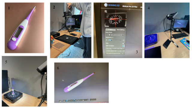
In the first picture you can see the digital thermometer that was used for the scanning. The first crucial step when using a 3D scanner is the calibration, which you can see being done in the second picture. Due to the reliance of light for the entire process, change in lighting can affect the quality of the scan. When calibrating you are allowing the device to calibrate to the current state of the surrounding light. In the third picture you can see the settings that are available before you scan. The brightness helps and determines what the scanner will be able to pick up. When you can see the shape of the object clearly and there are some red dots on the monitor, the brightness is adjusted well for the scan. The red represents extreme contrast. You adjust the brightness depending on the colors of the object being scanned. In the fourth picture you can see the 3D scanner mid scan. It displays purple laser lines vertically and flashes a white light and on the device. The fifth picture shows another position for the thermometer. It is important to get multiple angles when 3D scanning in order to get the best data from the device. The software is able to detect similar geometry and then combine the different scans together in order to create the best representation of the object. The sixth picture is what the final scanned 3D model looks like in the software, which can then be uploaded into a CAD program.