As students of the Honors class NHS Scotland- Policies, Problems, and Innovative Solutions, our study abroad program provided us the opportunity to tour the University of Dundee’s Center for Anatomy and Human Identification laboratory. Throughout the two-hour experience, we learned about the comprehensive and complex donation system, whereby many donors are local residents. The tour was segmented into three different parts. The first part of the tour was an introduction session where the head of the donations department discussed the responsibilities and policies followed by curators, researchers, and students to ensure the utmost respect is had when handling donors’ bodies. The second was an immersive exploration of the dissection zone; the third was an informative walkthrough of the storage spaces and preservation techniques.
This event was a first for me, like many other students in the program. While some students, such as myself, were more curious than others, I believe the situation had both positive and negative aspects. Cadaver donation is paramount to furthering our modern understanding of the body and its systems, whereby strides in learning can lead to innovation and a more well-rounded approach to healthcare. I found it extremely interesting and valuable to witness how medical students and researchers examine the body via cadavers. Comparatively, the experience required a great deal of emotional processing. Most of us have never seen a corpse in real life, thereby making the dissection aspect of it both rewarding but also challenging to evaluate. However, I am incredibly grateful to the program administrators at UNC-Chapel Hill and the University of Dundee for organizing the tour and the cadaver donors for giving us an opportunity that few our age are offered.
This experience changed my perspective about healthcare because, in retrospect, I learned how essential body donation is to improving healthcare. For medical students in training and degree-holding researchers/medical professionals, a wide array of skills, techniques, and ideas can be gained and created using a cadaver. As mentioned in the tour, med-students in residency can practice surgery before operating on a live patient, reducing the number of mistakes or errors in a real-time surgery. Alternatively, forensic pathologists can access parts of the body that enable them to better understand anatomy systems in ways that cannot be achieved using a living patient. Concerning biomedical engineering, a new product/part could be manufactured and outfitted to a cadaver before trials on a live patient to achieve the lowest levels of invasiveness or scarring on the body during insertion. In summation, I would highly recommend and strongly encourage prospective students of this UNC study abroad program to participate in this experience, as it was both eye-opening and enriching for someone interested in healthcare.
Author: mattpete
Peter Browns 3D Print & OnShape Reference [Technical Module Individual Assignment 1]
For this assignment, I created an OnShape design of an object related to healthcare and produced a physical model via 3D printing. I am fascinated by dentistry and oral health, making a toothbrush the perfect option. The measurements used for my OnShape design were true to scale; however, I had to decrease the size by twenty percent to print my toothbrush. Overall, this assignment was both challenging yet extremely rewarding as I gained new skills and insight into the logistics and elements required to design and 3D print an object.
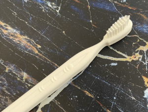
Top/Side view of printed model
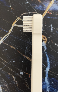
Side view of printed model
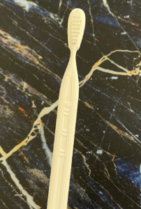
Front view of printed model
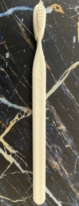
Front view of printed model (entire body)
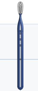
Front view of OnShape model (entire body)
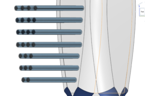
Bristle detail on OnShape
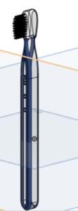
Side view of OnShape model (entire body)
Peter Browns 3D Print & OnShape Reference [Technical Module Individual Assignment 2]
Using Onshape, I designed an object that reflected my personal experience in Scotland. My object was a variation of the Loch Ness Monster. Nessie was printed in a size that allows him to be displayed on a desk, bookshelf, or even a car dashboard, wherever one may fancy! His creative design includes five separate parts that, when presented on a flat surface, make it appear as though he is breaching the surface of the water and gliding over Loch Ness. I selected this object because one of our first group activities following our arrival in Scotland was a hike around Loch Ness lake. Ultimately, it was one of my favorite experiences and memories from this study abroad trip!
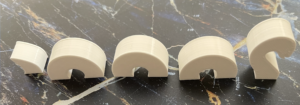
Top/Side profile view

Bottom/Side profile view
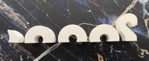
Side profile view
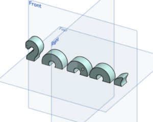
OnShape model design
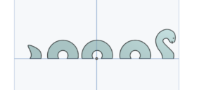
OnShape model design
Peter Brown’s Trip to Surgeons’ Hall
On Tuesday, June 28th, Sydney Schwartz and I visited the Surgeons’ Hall Museum in Edinburgh, Scotland. While we were interested in different exhibits, we both found the museum (as a whole) to be fascinating and incredibly eye-opening. The tour started in a central room that showcased tools used for surgeries dating back to the early 18th century (wow; we advanced in technology and innovation!). In the central room, many tumors and organs with abnormalities and several cadavers were on display. Two cadavers were the remains of children whose bodies were donated to science in 1702 and 1718 and were also the oldest in the museum’s collection. The cadaver from 1718 was dissected by Alexander Munro Primus, the first professor of anatomy at the Edinburgh University medical school.
One of the most interesting displays was a plaster cast of Robert Penman’s lower jaw tumor. While the tumor needed to be removed for Penman to sustain life (as he could not eat or drink due to the size of the tumor), the surgery was performed prior to the availability of effective anesthesia. Nonetheless, the surgery, performed by Dr. James Syme in 1828, was a success!
My favorite part of this museum visit was the dental surgery section. I especially enjoyed learning about early dental practices, particularly impression techniques. As one museum label explains, a dental impression is a negative imprint of teeth and mouth tissues that is used as a mound to make a positive cast. The tray on display would have been filled with soft material, like beeswax or plaster of Paris, to make the impression, as alginates or irreversible hydrocolloids had not yet been discovered as an effective/practical material.
The museum also featured modern technology and explained how it has improved healthcare. For example, biomedical engineering is an integral part of modern dentistry as virtual depictions of the jaw enable maxillofacial surgeons, dentists, and orthodontists to visualize vantage points inside their patient’s mouths for better accessibility.
The Surgeons’ Hall Museum was one of the highlights of my trip to Edinburgh. I would encourage anyone interested in medicine, healthcare, or technology (including biomedical engineering) to visit! No matter your specific curiosity, there is an exhibit in the museum sure to challenge your way of thinking concerning healthcare or the advancement of modern medicine.
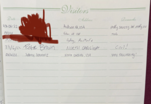
Our signatures in the visitors log
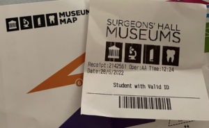
My ticket and museum brochure
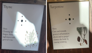
Early uses of natural remedies!
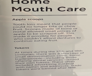
Interesting description about the limitations of missing teeth!
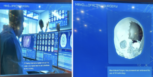
How biomedical engineering is used in Maxillofacial Surgery!
